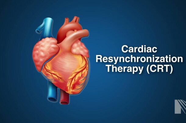
Cardiac resynchronization therapy (CRT) is a medical procedure that involves the implantation of a specialized device known as a biventricular pacemaker or cardiac resynchronization device. This therapy is typically used to treat certain types of heart conditions, such as congestive heart failure, where the heart’s chambers do not contract in a coordinated and efficient manner.
How CRT Works
1. Device Implantation: During the CRT procedure, a biventricular pacemaker is surgically implanted under the skin in the chest area. Wires or leads are threaded through veins and positioned in the right atrium, right ventricle, and left ventricle of the heart.
2. Electrical Signals: The biventricular pacemaker sends electrical signals to both the right and left ventricles of the heart. These signals are synchronized and timed to help coordinate the contraction of the ventricles.
3. Improved Coordination: By delivering electrical impulses to both ventricles, the CRT device helps the heart’s chambers contract in a more synchronized and efficient manner. This coordination improves the heart’s ability to pump blood and can be especially beneficial for individuals with heart failure or certain arrhythmias.
CRT is primarily used in cases where a patient’s heart’s electrical system is not functioning properly, leading to a lack of coordination in the heart’s contractions. This lack of coordination can result in symptoms like fatigue, shortness of breath, and fluid retention. By improving the heart’s pumping ability, CRT can help alleviate these symptoms and enhance the patient’s quality of life.
An implantable cardioverter-defibrillator (ICD) is a specialized medical device that is implanted in the chest, usually just below the collarbone. Its primary function is to monitor the heart’s rhythm continuously and deliver therapeutic interventions when it detects life-threatening arrhythmias. Here’s how an ICD works and when it’s typically recommended:
Monitoring the Heart: The ICD continuously monitors the electrical activity of the heart. It can detect various types of arrhythmias, with a focus on ventricular tachycardia and ventricular fibrillation, which are extremely fast and chaotic heart rhythms that can be life-threatening.
Electric Shocks: When the ICD detects a dangerous arrhythmia, it can deliver precisely calibrated electrical shocks to the heart. These shocks are intended to restore a regular heart rhythm. This process is known as defibrillation.
ICDs are recommended in the following situations:
History of Ventricular Arrhythmias: Patients who have experienced ventricular tachycardia or ventricular fibrillation in the past may be candidates for an ICD. These arrhythmias can lead to sudden cardiac arrest.
High Risk of Ventricular Arrhythmias: Even in the absence of prior arrhythmias, some individuals may be considered at high risk for developing them. This could be due to various factors, including a weakened heart muscle (as seen in heart failure) or certain genetic conditions.
Prior Heart Attack: After surviving a heart attack, some individuals are at an increased risk of ventricular arrhythmias, and an ICD may be recommended as a preventive measure.
Certain Genetic Conditions: Inherited cardiac conditions that predispose individuals to life-threatening arrhythmias, such as long QT syndrome, may also warrant the use of an ICD.
ICDs can significantly improve the survival rate for individuals at risk of sudden cardiac death due to ventricular arrhythmias. The device can provide a rapid response to these life-threatening events, effectively restoring a normal heart rhythm and preventing sudden cardiac arrest.
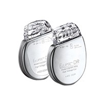
Ellipse ICDs
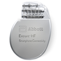
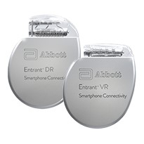
Entrant ICDs
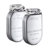
Fortify Assura ICDs
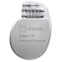
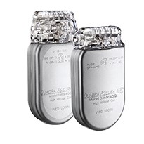
Gallant ICDs
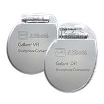
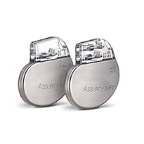
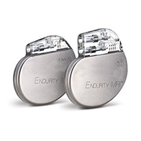
PACEMAKERS
A pacemaker is a small, battery-powered medical device that is surgically implanted to manage and regulate the heart’s rhythm. It is typically placed under the skin near the collarbone. Here’s a more detailed explanation of pacemakers and the different types:
Function of a Pacemaker: The primary function of a pacemaker is to monitor the heart’s electrical activity and, if necessary, send electrical impulses to the heart muscle to regulate its rhythm. This is particularly important for individuals with bradycardia, a condition where the heart beats too slowly.
Types of Pacemakers:
Single Chamber Pacemaker: This type of pacemaker typically has one lead (wire) that is placed in either the right atrium or right ventricle of the heart. It sends electrical signals to the lower right chamber of the heart to help maintain a regular heartbeat.
Dual Chamber Pacemaker: In a dual chamber pacemaker, there are two leads—one in the right atrium and one in the right ventricle. This allows the device to send electrical signals to both the upper and lower right heart chambers. Dual chamber pacemakers are used to help coordinate the timing of atrial and ventricular contractions more effectively.
Biventricular Pacemaker (Cardiac Resynchronization Pacemaker): This type of pacemaker is typically used for individuals with heart failure and a condition known as left bundle branch block, which causes asynchronous contractions of the heart’s ventricles. A biventricular pacemaker has three leads—one in the right atrium, one in the right ventricle, and a third lead placed in the left ventricle. By stimulating both lower heart chambers (ventricles), it aims to improve the synchronization of heart muscle contractions and, in turn, enhance the heart’s pumping efficiency.